How do we prepare our meticulously crafted DNA libraries for the high-tech world of nanopore sequencing, ensuring every molecule is ready to tell its story?
The journey of metagenomics involves many intricate steps, from initial sample collection to DNA extraction and barcoding. But even after isolating and tagging our DNA fragments, they aren’t quite ready for the sequencer. This final preparation stage is crucial, ensuring our DNA library can interact seamlessly with the advanced nanopore technology, leading us from the wet lab to the fascinating realm of bioinformatics.
Assembling the Sequencing-Ready Library
After the barcoding and pooling steps, the library needs sequencing adapters. These specialized DNA sequences are essential for the nanopore system to recognize, capture, and process each individual DNA molecule as it passes through the tiny pores.
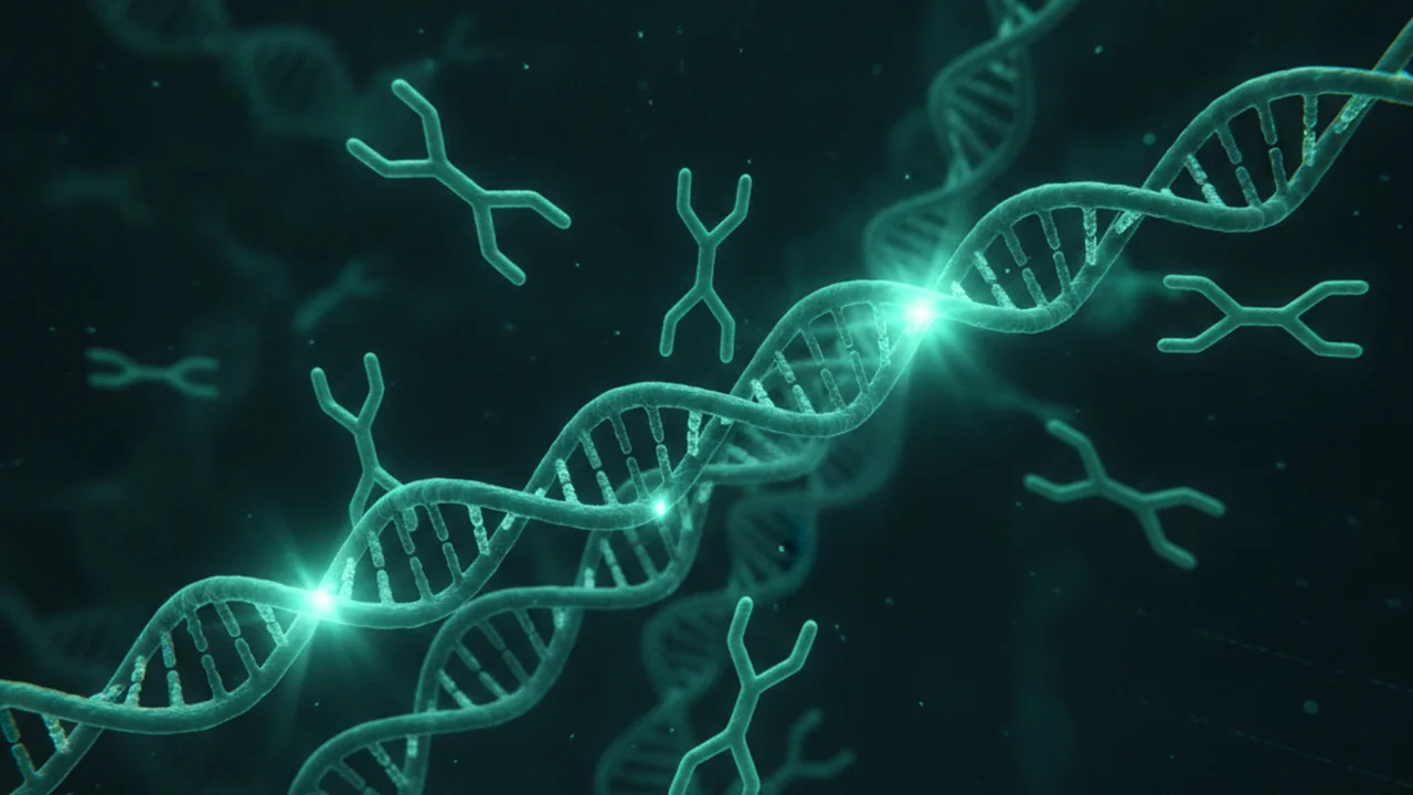
Short, synthetic DNA sequences ligated to the ends of DNA fragments in a library. They contain essential functional elements for sequencing, such as binding sites for the sequencing platform, primer binding sites, and unique molecular identifiers.
The Adapter Ligation Reaction
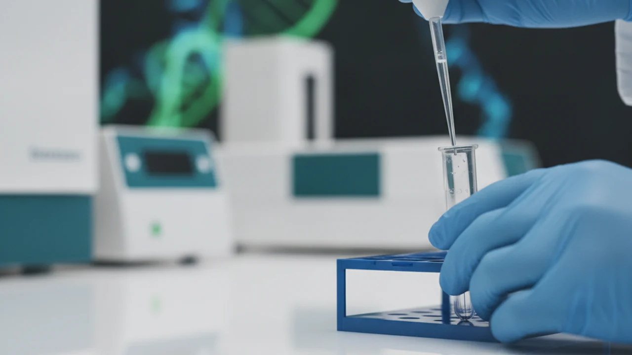
In the adapter ligation reaction, the pooled barcoded sample is mixed with adapter mix, ligation buffer, and Quick T4 DNA ligase. This enzyme attaches adapters to the prepared DNA ends, forming stable phosphodiester bonds that integrate the adapters into the DNA library.
Want to go deeper? The science behind T4 DNA Ligase…
T4 DNA ligase is an enzyme isolated from the T4 bacteriophage. It catalyzes the formation of a phosphodiester bond between adjacent 5′-phosphate and 3′-hydroxyl termini in DNA. In adapter ligation, the enzyme requires ATP as a cofactor to create a high-energy adenylated intermediate, which then facilitates the sealing of nicks in double-stranded DNA, effectively joining the adapters to the library fragments. Its efficiency and specificity are critical for successful library preparation.
Cleanup and Elution: Refining the Library
A final bead cleanup removes unligated adapters and reaction components. The DNA library is washed multiple times using Long Fragment Buffer, briefly dried, and then eluted into a small volume of buffer (typically 15 microliters elution buffer). This purification step is crucial to ensure a clean, concentrated library, free from contaminants that could hinder sequencing.
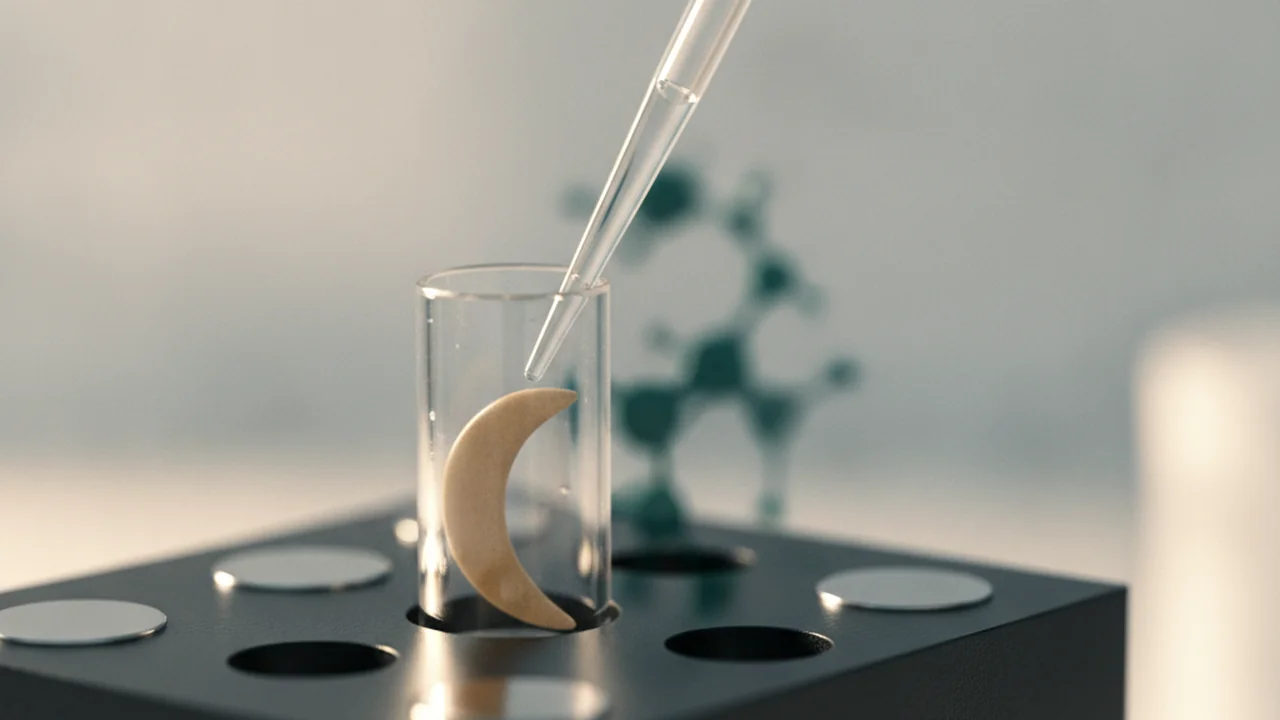
Unligated adapters can compete with the prepared library for binding sites on the flow cell, leading to reduced sequencing efficiency and an abundance of adapter-only reads, which are costly to process and provide no biological information.
Imagine you’re designing a new cleanup protocol. How would you visually represent the successful removal of unwanted components and the retention of your desired DNA library?
- Draw a simple diagram showing a microtube before and after bead cleanup.
- Label the DNA library, unligated adapters, and other reagents.
- Indicate how the beads and washes help achieve purification.
Consider the impact of even small contaminants on the overall success of a sequencing experiment.
Quantifying the Library: The Qubit Fluorometer
The final library is quantified using a Qubit fluorometer. This highly sensitive method measures double-stranded DNA specifically. The protocol recommends loading about five to ten femtomoles of prepared library onto the flow cell. Accurate quantification is essential to optimize pore occupancy and maximize the data yield from your sequencing run.
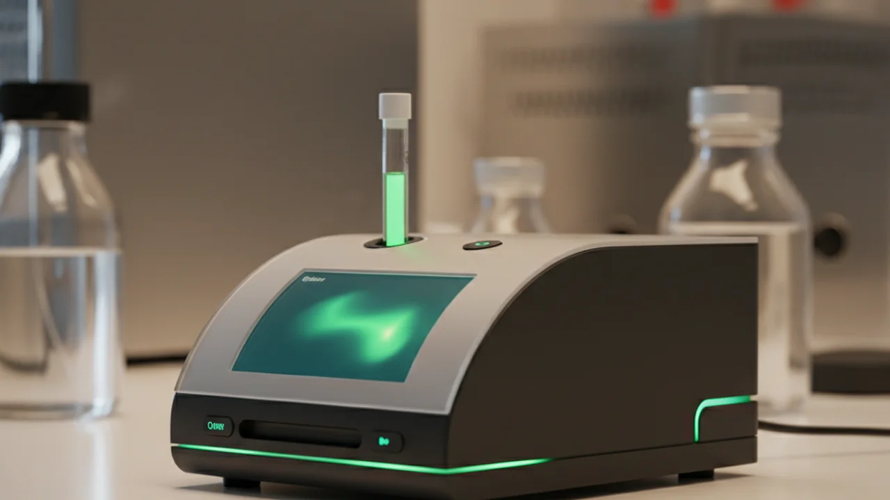
Measuring DNA concentration with a spectrophotometer (e.g., NanoDrop) is sufficient for sequencing library quantification.
Spectrophotometers measure all nucleic acids, including RNA and free nucleotides. A fluorometer like Qubit uses fluorescent dyes specific to double-stranded DNA, providing a much more accurate and reliable concentration for sequencing libraries, which is critical for consistent results.
“Once sequencing begins, the work shifts from wet lab technique to bioinformatics: turning raw signal into biological meaning.”
Introducing the Nanopore Device: The MinION
With the library quantified, our focus shifts to the sequencing platform itself. Oxford Nanopore sequencing reads DNA as molecules pass through tiny nanopores. Changes in electrical signal are converted into sequence data, providing real-time insights into the genetic material.
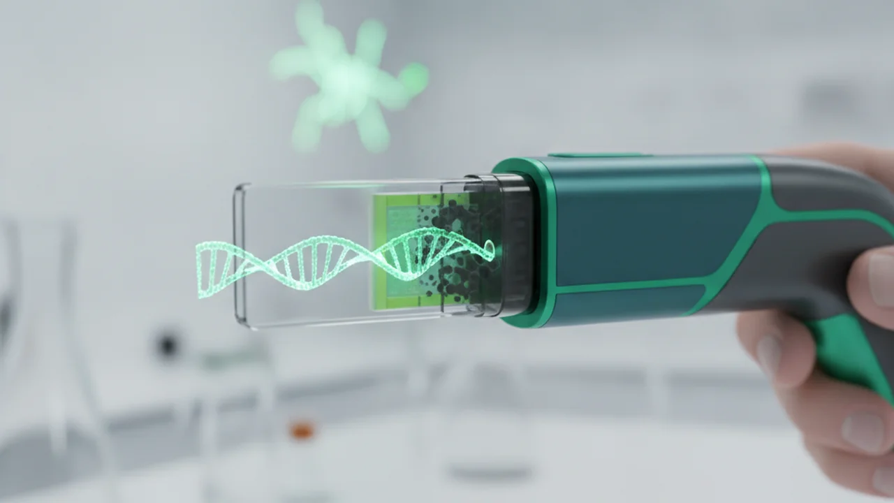
Nanopore sequencing is revolutionizing fieldwork, enabling real-time pathogen detection, environmental monitoring, and even space biology experiments due to its portability and rapid data generation.
Flow Cell Check and Priming: Preparing for Sequencing
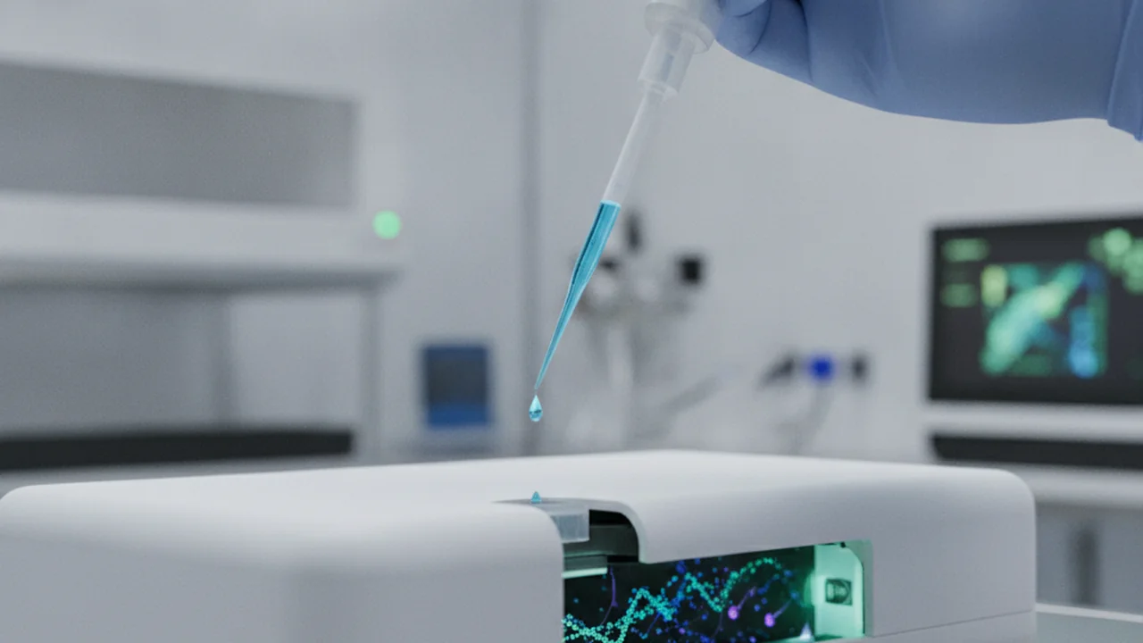
Before loading the library, the flow cell must be checked and primed. This involves careful handling to protect the delicate pores and precisely pipetting buffer into the priming port. This prepares the fluid path for sequencing, ensuring the library can efficiently access the nanopores.
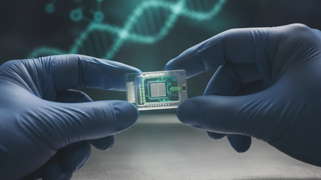
Reflect on the importance of meticulousness in laboratory procedures like flow cell priming. How does attention to detail at each step contribute to the overall reliability and success of a complex experiment?
Loading the Library and Post-Sequencing Wash
The final library is loaded onto the flow cell, initiating the sequencing run. After sequencing, washing helps clear the flow cell and manage the waste channel according to the workflow. This ensures that the flow cell is clean and ready for potential re-use or proper disposal.
- Sequencing adapters are crucial for nanopore recognition and processing.
- Adapter ligation uses T4 DNA ligase to attach adapters to DNA fragments.
- AMPure bead cleanup removes contaminants for a pure, concentrated library.
- Qubit fluorometry provides accurate DNA quantification for optimal flow cell loading.
- Flow cell priming and careful handling are vital for successful sequencing.
How do the wet lab techniques discussed here directly influence the quality and quantity of data obtained from sequencing?
From Wet Lab to Bioinformatics: Bridging to Data Analysis
Once sequencing begins, the work shifts from wet lab technique to bioinformatics: turning raw signal into biological meaning. Device signal traces become FASTQ files and are visualized on software dashboards. This transition marks the shift from physical sample manipulation to computational analysis, where the real stories within the metagenome begin to unfold.
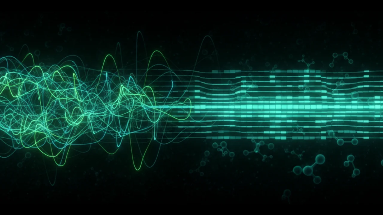
What is the primary output format when raw electrical signals from nanopore sequencing are converted into sequence data?
Successful nanopore sequencing hinges on meticulous library preparation, including adapter ligation, thorough cleanup, and accurate quantification, followed by careful flow cell handling and the critical transition to bioinformatics for data interpretation.
What is the main purpose of sequencing adapters in nanopore sequencing?
The Shift
- The final stages of library preparation, including adapter ligation and rigorous cleanup, are paramount for successful nanopore sequencing, ensuring DNA fragments are correctly recognized and processed.
- Accurate quantification of the DNA library, typically with a Qubit fluorometer, is critical to optimize flow cell loading and maximize sequencing output.
- The process transitions from precise wet lab techniques to sophisticated bioinformatics, where raw electrical signals are converted into meaningful biological data for interpretation.